Home
Uncategories
Smooth Muscle Diagram - Schematic Diagram Of The Regulation Of Smooth Muscle Contraction Gpcr Download Scientific Diagram / The cardiac muscle is only found in the heart wall.
Smooth Muscle Diagram - Schematic Diagram Of The Regulation Of Smooth Muscle Contraction Gpcr Download Scientific Diagram / The cardiac muscle is only found in the heart wall.
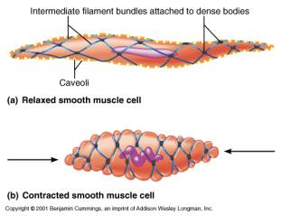
Smooth Muscle Diagram - Schematic Diagram Of The Regulation Of Smooth Muscle Contraction Gpcr Download Scientific Diagram / The cardiac muscle is only found in the heart wall.. The smooth muscles perform the functions in the contrast of other types of muscles. Smooth muscle is a type of muscle tissue which is used by various systems to apply pressure to vessels and organs. Smooth muscles exhibits a phenomenon called _____ in which: The calcium is the cause of protein to detach from the actin and myosin fastly binds with the opening of actin. Smooth muscle tissue, unlike striated muscle, contracts slowly and automatically.
Smooth muscle is a type of tissue found in the walls of hollow organs, such as the intestines, uterus and stomach. Drawing exercise sectional smooth muscle tissue labeled diagram | world of reference. Diaphragm is also a skeletal muscle. Smooth muscle is composed of sheets or strands of smooth muscle cells. It is the pen diagram of skeletal, smooth and cardiac muscle for class 10, 11 and 12.
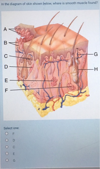
Solved In The Diagram Of Skin Shown Below Where Is Smoot Chegg Com from media.cheggcdn.com Diaphragm is also a skeletal muscle. Smooth muscle makes up the walls of hollow organs, respiratory passageways, and blood vessels. The cells stick together and are connected by specialised cell junctions, called gap junctions. Transcribed image text from this question. Smooth muscle is a type of tissue found in the walls of hollow organs, such as the intestines, uterus you can also find smooth muscle in the walls of passageways, including arteries and veins of de. Smooth muscle has a fusiform shape, which resembles a football or spindle. Smooth muscle tissue is also known as visceral muscle tissue. Following is a diagram of the diaphragm :
This smooth muscle can be found surrounding the walls of the blood vessels, the bronchioles in the lungs, and the sphincter muscles used in the gi tract.the gi tract, which is tubular by design, also houses longitudinal muscles in addition to the smooth.
This figure shows the structure of the muscle fibers. Smooth muscle (textus muscularis levis) smooth muscle is a type of tissue found in the walls of hollow organs, such as the intestines, uterus and stomach. Cardiac, skeletal and smooth muscles are the three types of muscles found in the human body. Arteries have thick walls due to smooth muscle cells, which help them carry blood away from the heart to every part of. Beta 2 receptors are also on small coronary arterioles thus increasing hormonally induced blood flow within the musculature of the heart. Smooth muscle determines the flow of blood in the arteries. It constitutes much of the musculature of Smooth muscle cell labeled diagram ~ diagram. Muscle anatomy ankle 12 photos of the muscle anatomy ankle ankle anatomy muscle tendon, foot ankle. Related posts of smooth muscle diagram muscle anatomy ankle. Smooth muscle lines the inside of blood vessels and organs, such as the stomach, and is also known as visceral muscle. Smooth muscle is found in the walls of hollow organs like your intestines and stomach. Smooth muscle anatomy smooth muscle tissue is also known as visceral muscle tissue.
Smooth muscle is found in the walls of hollow organs like your intestines and stomach. Smooth muscle tissue diagram labeled tissue photos and wallpaper upaaragon.co. Also, after activation of the receptors there is a long process in order to elicit an action potential, involving second. • smooth muscles respond to stretch only briefly, and then adapts to its new length • the new length however, retains its original _____ seconds or minutes after it has been elongated or shortened (e.g. Smooth muscles in arteries and veins are largely responsible for regulation of blood pressure.
Histology Of Muscle from faculty.etsu.edu Here's a quick rundown of the key. It is the pen diagram of skeletal, smooth and cardiac muscle for class 10, 11 and 12. In this video i have shown the simplest way of drawing muscle drawing. Diagram of smooth muscle contraction, smooth cardiac and skeletal muscle diagram, smooth muscle cell diagram, smooth muscle cell picture. It is layered in a distinctive pattern of circular layers. The cells stick together and are connected by specialised cell junctions, called gap junctions. You will have some basic understanding of the appearance referring to the below smooth muscle diagram. It is the pen diagram of skeletal, smooth and cardiac muscle for class 10, 11 and 12.
Smooth muscles in arteries and veins are largely responsible for regulation of blood pressure.
It is the weakest type of muscle but smooth muscles in the gastrointestinal or gi tract control digestion. It is the pen diagram of skeletal, smooth and cardiac muscle for class 10, 11 and 12. The cells stick together and are connected by specialised cell junctions, called gap junctions. Draw a diagram of smooth muscle fibre and label any three parts. Back muscle chart 12 photos of the back muscle chart back muscle diagram human body, back muscle diagram pain, back muscle groups diagram, back muscle workout diagram, lower back muscle chart, human muscles, back muscle diagram human body, back muscle diagram pain, back muscle groups diagram, back muscle workout diagram. Circuit diagram used for study 1141x1080 draw the diagram of smooth muscles or neuron muscle smooth muscle is found in the walls of hollow organs like your intestines and stomach. Also, after activation of the receptors there is a long process in order to elicit an action potential, involving second. The smooth muscle contraction is much slower than in the striated muscle primarily due to the presence of g protein coupled ligand receptors instead of ion channel coupled ligand gated receptors present in striated muscle. Drawing exercise sectional smooth muscle tissue labeled diagram | world of reference. In skeletal muscle, a single type of somatic nervous system traverses to muscle, where it stimulates organelle in the muscle cells in order to release calcium. Smooth muscles in arteries and veins are largely responsible for regulation of blood pressure. Beta 2 receptors are also on small coronary arterioles thus increasing hormonally induced blood flow within the musculature of the heart. This figure shows the structure of the muscle fibers.
It constitutes much of the musculature of It is the pen diagram of skeletal, smooth and cardiac muscle for class 10, 11 and 12. Related posts of smooth muscle diagram labeled back muscle chart. Smooth muscle has a fusiform shape, which resembles a football or spindle. The cells stick together and are connected by specialised cell junctions, called gap junctions.
Overview Of The Muscular System Boundless Anatomy And Physiology from textimgs.s3.amazonaws.com It is the pen diagram of skeletal, smooth and cardiac muscle for class 10, 11 and 12. Smooth muscle tissue is also known as visceral muscle tissue. Related posts of smooth muscle diagram muscle anatomy ankle. Also, after activation of the receptors there is a long process in order to elicit an action potential, involving second. In skeletal muscle, a single type of somatic nervous system traverses to muscle, where it stimulates organelle in the muscle cells in order to release calcium. Smooth muscle tissue, unlike striated muscle, contracts slowly and automatically. The cardiac muscle is only found in the heart wall. Smooth muscle has a fusiform shape, which resembles a football or spindle.
The cardiac muscle is only found in the heart wall.
Smooth muscle makes up the walls of hollow organs, respiratory passageways, and blood vessels. The cardiac muscle is only found in the heart wall. This is just a diagram of how the human muscle looks under all the tissue and skin. This smooth muscle can be found surrounding the walls of the blood vessels, the bronchioles in the lungs, and the sphincter muscles used in the gi tract.the gi tract, which is tubular by design, also houses longitudinal muscles in addition to the smooth. Smooth muscle is found in the walls of hollow organs like your intestines and stomach. It is the pen diagram of skeletal, smooth and cardiac muscle for class 10. For example muscles of limbs. The smooth muscle contraction is much slower than in the striated muscle primarily due to the presence of g protein coupled ligand receptors instead of ion channel coupled ligand gated receptors present in striated muscle. Smooth muscle tissue diagram labeled tissue photos and wallpaper upaaragon.co. Smooth muscle is a type of muscle tissue which is used by various systems to apply pressure to vessels and organs. It is layered in a distinctive pattern of circular layers. Smooth muscle diagram | quizlet. You will have some basic understanding of the appearance referring to the below smooth muscle diagram.

0 Comments:
Posting Komentar